IP Screening
1. Prepare an appropriate set of extraction solvents(better be fresh- equilibrated to RT for extraction and then at 4°C (or held on ice)) : 2.2 ml of each solution in the wells of a 2.5 ml 96-well deep-well microplate (SOLVENT PLATE) and set aside.
2. Prepare a plate containing 2 μl of 500 mM DTT in each appropriate well of a 250 μl 96-well PCR plate (LOADING PLATE).
3. Prepare a 0.8 ml 96-well deep-well microplate with 100 μl of each extraction solution (SOLVENT PLATE) in the relevant wells. Dispense 5 μl of magnetic affinity medium slurry (mix well) in each of well (BINDING PLATE), we used another batch of flag beads for the last 7 wells. Wash the beads in each well by moving the plate on the magnet. Place the plate on the deep-well magnet, remove the washing solutions and hold covered at 4°C until ready for use, stable for several hours.
4. Prepare protease inhibitors by dispensing 5 μl of 100x stock into each of 24 wells of a 0.8 ml 96-well deep-well microplate (PI PLATE), store frozen until use. These inhibitors will be included in the procedure by combining with extraction solvent and transferring the mix to frozen cell powders.
5. Ensure that the SOLVENT PLATE and PI PLATE are equilibrated to RT. Dispense an appropriate amount of cell powder to each well, using a dispensing manifold or using a volumetric spoon. Pre-cool the manifold setup with liquid nitrogen (LN2). Cell powder should also be cooled on LN2. Dump cell powder onto the manifold surface and pack it into all the wells of the manifold using the packing tool. Excess powder is recovered using a spatula. A pre-cooled 0.8 ml 96-well deep-well microplate (EXTRACTION PLATE) is placed on top of the manifold such that the openings of the wells of each device are aligned and face each other; and then the sandwich is inverted, spacers are removed, and it is given a firm tap. Proteins in the ~10,000+ copies per cell abundance range, ~35-50 mg (37mg STAT3 3XFLAG cells were used) of human cell powder can provide for robust colloidal Coomassie blue G-250 staining35 of all major components of an enriched mixture after IP (yields in the tens to hundreds of ng per band).
6. Remove the EXTRACTION PLATE from LN2 and allow it to stand ~2 minutes at RT. The powders will remain thoroughly frozen during this time. Add 450 μl (for 50 mg human cell powder) of each RT solvent from the SOLVENT PLATE to the PI PLATE – producing solvents including 1x protease inhibitors. Transfer this entire mixture to the concordant wells of the EXTRACTION PLATE. Cover the plate with a cap mat that has had the caps from the appropriate columns pierced to allow the use of a multi-microtip probe sonication and transfer the plate to 4°C.
7. For ~37 mg of human cells, sonication is carried out with an 8-microtip probe using an amplitude setting of 1. Apply sonication to one row (250 J total/row).
8. Using an 8-channel pipette with adjustable channel width, transfer crude extracts from the EXTRACTION PLATE to 24 x 1.5 ml tubes and centrifuge for 10 min, ~20,000 RCF at 4 °C in a bench top microcentrifuge.
9. Transfer the supernatants, one-at-a-time, using a standard pipette to the BINDING PLATE and cap the plate with a cap mat or other liquid-tight plate seal. Resuspend the beads fully within each well by manually inverting the plate several times. Place the plate on a rotating wheel at moderate speed (prevent the beads from settling) for 30 min – 1 hr.
10. Remove the BINDING PLATE from the rotating wheel and briefly spin down to collect all liquid within the bottom of the well: e.g., 1-2 min at 2k RPM and 4°C.
11. Place the BINDING PLATE on a 96-well deep-well magnet to retain the beads at the well sides. Then remove the depleted extracts and remove the plate from the magnet. After removing the solution, remove the plate from the magnet.
12. Add 500 μl of concordant extraction solvent to each well of the BINDING PLATE (wash #1). Place the plate back on the magnet and alter its position several times to wash and mix. Park the plate back in the original position on the magnet to collect and hold the beads at the well side and remove the wash. Then remove the plate from the magnet. 13. Repeat Step 12 (wash #2), then proceed to Step 14
14. Add 200 μl of concordant extraction solvent to each well of the BINDING PLATE (wash #3). Pipette the solution in each well up and down several times to fully resuspend the beads and transfer them to a 250 μl 96-well PCR plate (ELUTION PLATE) placed on a 96-well PCR plate magnet. Removing the wash solution once the beads have adhered to the well sides.
15. Briefly spin down (as in Step 10) and place the ELUTION PLATE on the 96-well PCR plate magnet to remove any remaining wash solution from the prior step. 16. Add 18 μl of 1.1x LDS loading buffer to the sample. Cover with a strip tube caps or a plate seal and incubate the plate 5- 10 min at between RT and 70°C with vigorous shaking. Re-equilibrate the plate to RT if heated. Briefly spin the plate to recapture all beads and loading buffer at the bottom of the wells as in Step 10 (operation at RT is fine). Place the ELUTION PLATE on the 96-well PCR plate magnet and transfer the elutions to a plate containing 2 μl 500 mM DTT (LOADING PLATE).
17. Cover the sample-containing wells of the plate with strip caps or thermal seal and reduce the samples by heating to 70°C for 10 min. Cool to RT. Briefly spin the plate to recapture all the solutions at the bottom of the wells.
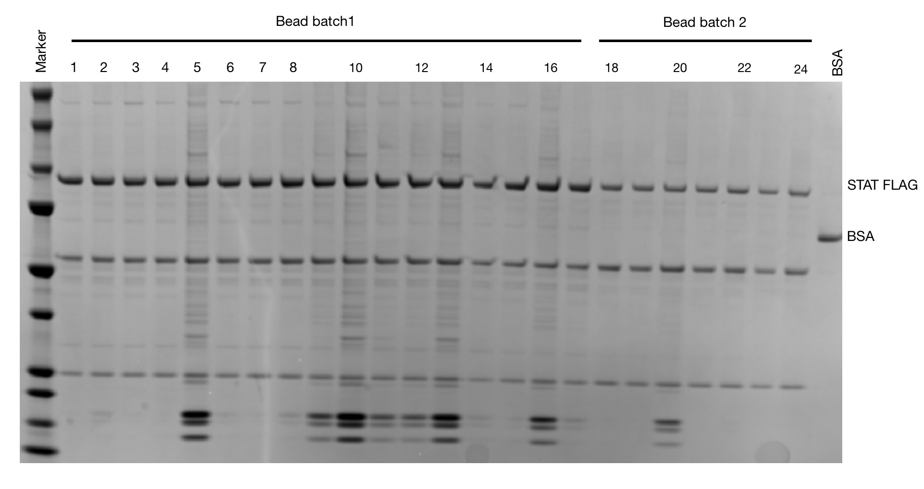
18. Load all samples on a 26-well 4-12% Bis-Tris midi-gel and stain with silver blue with marker and 50ng BSA.